
The Intricate World of Mold: A Look at Hyphae Filaments Under the Microscope
Introduction to Mold Filaments
Mold is a fascinating kingdom of fungi that plays a vital role in our ecosystem. At the heart of this microscopic world are hyphae filaments, which are the essential building blocks of mold organisms. These delicate structures not only contribute to the growth and reproduction of molds but also interact with their environments in complex ways. This blog post aims to explore the structure and function of hyphae filaments as observed under the microscope, providing insights into their remarkable features.
The Structure of Hyphae Filaments
Hyphae filaments are elongated structures composed of a series of cells linked together. Each filament can branch out to form a mycelium, which is the vegetative part of the mold. Under a microscope, these filaments display a characteristic appearance that varies between different mold species. Some hyphae are septate, meaning they are divided into compartments by internal cross-walls. Others, known as coenocytic hyphae, lack these divisions, forming a continuous tubular structure. This fundamental structural difference plays a significant role in their growth and nutrient absorption capabilities.
Function and Importance of Hyphae
The primary function of hyphae filaments is to absorb nutrients from their surroundings. As saprophytic organisms, molds degrade organic matter, recycling nutrients back into the ecosystem. Hyphae release enzymes that break down complex substances, allowing the mold to absorb simpler molecules. This process is crucial not only for the survival of molds but also for maintaining soil health and promoting plant growth.
Moreover, hyphae play a pivotal role in the formation of symbiotic relationships. For example, mycorrhizal fungi extend their hyphae into the root systems of plants, enhancing their access to water and essential nutrients. In return, molds receive organic compounds produced by the plants during photosynthesis. This mutualistic relationship highlights the essential nature of hyphae in ecological balance.
Visualizing Hyphae Filaments
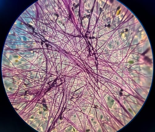
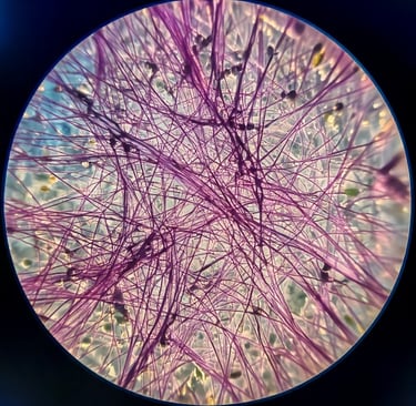
Observing hyphae filaments under a microscope reveals the beauty and complexity of these structures. With proper staining techniques, researchers can distinguish between different types of hyphae and gain insights into their functional roles. Microscopic imaging provides a clearer understanding of how molds interact with their environment, aiding in the study of biodiversity and the impacts of fungi on ecosystems.
In conclusion, hyphae filaments are more than mere structures; they are vital components of the mold kingdom with significant ecological implications. By studying these filaments under a microscope, we can appreciate the intricate relationships that fungi maintain within their environments. Understanding the biology of hyphae not only enriches our knowledge of the fungal world but also emphasizes the importance of these microorganisms in nurturing our planet's health.